
Public Titles
Mullerian duct anomalies (MDAs) are relatively common disorders that are particularly responsible for obstetrical complications and have a prevalence of about 3%–4% in the general population (1, 2).
Uterine malformations may result from arrested development of the mullerian ducts, failure of fusion of the mullerian ducts or failure of resorption of the median septum (1, 2).
Several classification systems have been used to describe MDAs. The most accepted system is the American Fertility Society (AFS) classification system. This classification is based on the embryologic etiologies behind MDAs. In this classifycation, class I consist of hypoplasia and agenesis of the uterus. Class II consists of unicornuate uterus. Class III is composed of uterus didelphys and class IV is characterized by bicornuate uteri. Class V anomallies are made up of septate uterus. Class VI includes an arcuate uterus and class VII is diethylstilbestrol-related anomaly (3, 4).
Accurate characterization of MDAs is crucial to the treatment process. As a result, screening for uterine anomalies forms a part of routine clinical investigations of woman with a history of infertility, recurrent miscarriages and early preterm labors (5). Various imaging modalities have been used in the evaluation of MDAs. Despite being invasive, hysterosalpingography (HSG), hysteroscopy and laparoscopy are the conventional methods for the assessment of uterine morphology.
Technologic advances in imaging modalities have revolutionized the evaluation of MDAs by noninvasive tools such as two-dimensional (2D US) and three-dimentional ultrasounds (3DUS) and MRI.
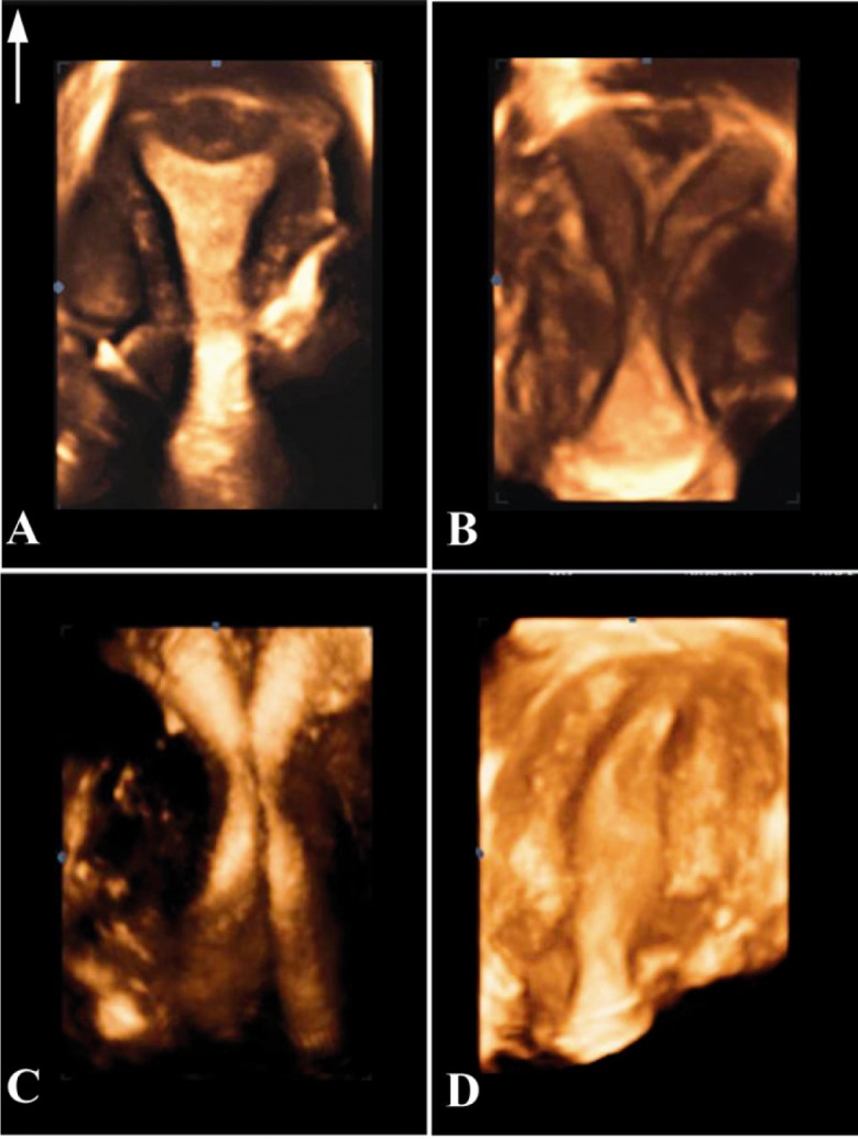
Despite the fact that two-dimensional sonography is in routine use because of its flexibility and moderate costs, but it has some limitations. The sensitivity of 2DUS especially for the demonstration of fundal contour is relatively low compared with other methods (6). On the other hand, 3DUS is a valuable tool for the assessment of uterine morphology because of its increased spatial awareness and the ability to visualize the coronal plane. The coronal view depicts both endometrial cavity and the serosal surface of the uterine fundus. Therefore, this view is a valuable problem-solving tool helping differentiate between various MDAs, including bicornuate, septate, unicornuate, and didelphys. Moreover, data acquisition time is short and images can be stored for later evaluation and analyzed as many times as needed (7).
In conclusion, endovaginal three-dimensional ultrasonography (3DUS) is a non-invasive, outpatient diagnostic modality, which enables a detailed assessment of the uterine morphology. Because of the high level of agreement between 3D ultrasonography and hysterosalpingography, MRI, hysteroscopy and laparoscopy, 3DUS has recently become the only mandatory step in the initial investigation of MDAs before resorting to invasive procedures such as hysteroscopy.
