
During early foetal life, cells that will develop into primary oocytes become rapidly enclosed by a subpopulation of somatic cells. These cells will be required to meet the demands of the growing oocytes during their development. Although primary oocytes can remain quiescent for years and are arrested in meiotic prophase I, a small percentage of them are recruited throughout life, even during pregnancy and non-ovulation periods to sustain the reproductive function (1). After resumption of meiotic maturation, changes in both somatic and germ cell lines take place. The dominant follicle will contain the oocyte destined to be ovulated, which will progress through meiosis until it arrests for the last time at the metaphase II (MII) stage waiting to be fertilized by a sperm whilst the other recruited follicles will undergo atresia. Concomitantly, somatic cells differentiate to different populations, but some (Cumulus cells (CCs)) will remain in close physical contact with the oocyte.
The presence of transzonal projections emerging from CCs along follicle development establishes a two-way cross talk and interactions with the oocyte. This biological communication between cell lines triggered the interest in the study of CCs as a biomarker of oocyte competence. Although research in transcriptomics has proven to be an exciting field with the potential to understand oocyte biology (2–4), there is currently a lack of consensus on the gene(s) that could serve as potential biomarkers in CCs. Currently, there is limited evidence on the use of CCs to determine germinal aneuploidy status. This case provides guidance to focus on proper follow-up investigations when a different cell line in a female patient is observed. This case report is about using the current gold standard next generation sequencing (NGS) combined with conventional karyotyping techniques to evaluate the underlying genetic status of an individual.
A 37-year-old single woman presented to the Centre for Reproductive and Genetic Health, London (UK) on 14th August 2019 for fertility cryopreservation. Ethical approval (10/H0709/26) and patient written consent was obtained in relation to this publication. She had no previous history of pregnancy or family history of premature menopause. Her medical history was unremarkable and her body mass index was 27.83 kg/m2 (Normal). Her ovarian reserve test revealed an anti-Mullerian hormone of 3.9 pmol/l and follicle stimulating hormone of 7.0 IU/L. The patient underwent three cycles of controlled ovarian stimulation to optimize the yield of oocytes. An antagonist protocol was prescribed with Menopur® 450 IU for both cycles. Vaginal oocyte retrieval was carried out 37 hr post Gonasi® (10.000 IU) trigger and a total of six oocytes were collected in the first cycle. Removal of cumulus cells was performed 39–41 hr post trigger by exposure to cumulase (Origio Specialty Pharma, Denmark). Four oocytes were mature (MII) and two were found to be at the germinal vesicle (GV) stage. Cumulus cells were transferred into a HEPES media with micro-drops overlaid with oil and used for training purposes to optimize a technique of cell amplification. Different concentrations containing three, four, six and eight CCs and a blank were placed into microcentrifuge tubes containing BSA/PBS buffer. Amplification was successful in three out of the four samples after next generation sequencing (NGS) using Ion ReproSeqPGS kits (Thermo Fisher Scientific Inc., US) (Figure 1). The samples containing four and six CCs showed a 45,X complement in the NGS profile whilst the one with eight cells showed 40–50% mosaicism for the same chromosomal aberration.
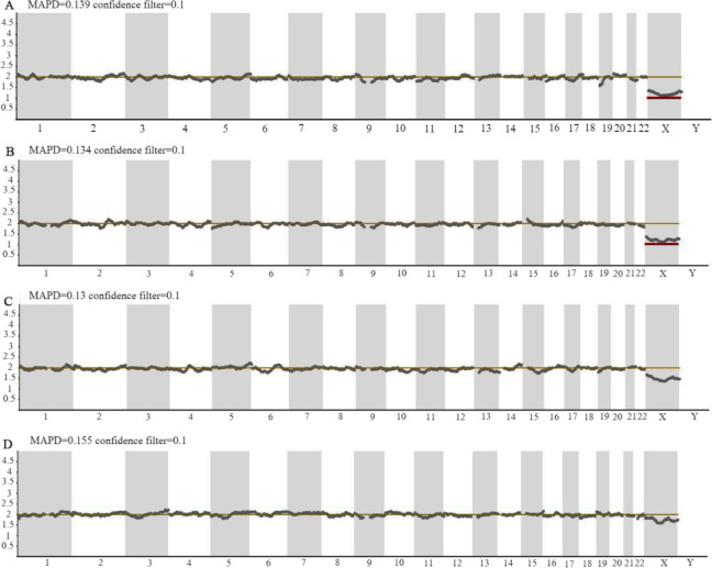
The patient was informed of the findings and a karyotype was strongly recommended for further clarification, since the patient had presented with low ovarian reserve parameters. An apparently normal female chromosome complement (46,XX) and banding pattern was noted after standard analysis on 5 metaphase lymphocyte cells (Supplemental Figure 1). In view of the referral indication, an extended screening of sex chromosomes was performed on a total of 50 metaphase cells where in two cells (4%), a complement of 45 chromosomes was seen with a single cell copy (Monosomy) of the X chromosome and in 48 (96%) cells, an apparently normal female chromosome complement was observed. A second cycle was performed following the same protocol resulting in five oocytes namely four MII stage and one metaphase I (MI) stage oocytes. The immature MI oocyte was subjected to the same NGS protocol as previously done on the cumulus cells in order to understand the origin of the mosaicism detected in the CCs. A third cycle was performed resulting in six oocytes, four MII stage, one MI and one GV. Immature oocytes were subjected to NGS protocol. A normal chromosome complement was noted in the immature oocytes (Supplemental Figure 2). In order to investigate the extent of somatic/germline mosaicism, a buccal swab was requested and sent for further NGS analysis. Extracted DNA from epithelial cells was diluted to 40 pg/μl and 2.5 μl were subjected to NGS. Results showed a 21% mosaicism for the 45,X complement (Figure 1). Results were conveyed to the patient and genetic counselling was provided to explain the implications of these findings and the repercussion on her fertility journey.
The presence of CCs with Turner Syndrome (TS) genotype led the team to refer the patient to a blood chromosome study. According to a study by Russell et al., for a patient aged 37 years, the reportable range for X chromosome loss within peripheral blood cultures would be 6% or above (5). At the observed level of 4%, the laboratory could not differentiate between genuine mosaicism, age related X loss or technical artefact. Interestingly, a previous report in literature has shown a patient diagnosed with non-mosaic TS following karyotyping of 50 lymphocyte cells being re-diagnosed as mosaic TS after observing a larger number of cells from different tissues using FISH (6). Hence, the observation of higher number of blood lymphocytes could show various genotype patterns.
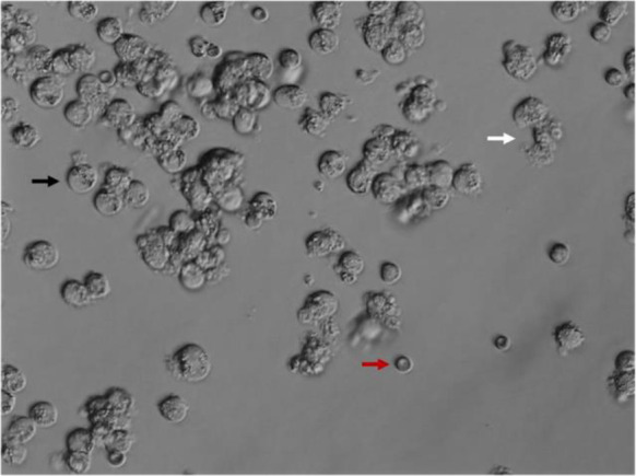
The detection of 45,X cells in a different tissue sample (Somatic cumulus cells) was initially done in our patient with no apparent abnormal phenotype. Although the differentiation of age-related loss versus genuine mosaicism is challenging, discovering aneuploid cells in a second somatic tissue (Buccal epithelial cells) would favor the latter. A normal female chromosome complement was noted in one metaphase I oocyte. A recent study including ovarian follicles from 5 female individuals with TS revealed that some oocytes were found to be normal after X chromosome analysis although granulosa cells were largely monosomic (7). Hematopoietic stem cells such as lymphocytes arise from the lateral mesoderm and granulosa cells are derivates from the intermediate mesoderm (8). Given that germ cells (Oocytes) also have their origin in the intermediate mesoderm, a certain degree of mosaicism could be expected. Peek et al. (9) also noted 45,X oocytes in human, although it has been highlighted in literature that oocytes may escape meiotic sex chromosome inactivation at higher frequency than spermatocytes (10). From the present case study, the validity of CCs as a tool for oocyte aneuploidy can be confirmed. However, validity of CCs chromosomal analysis as a predictive tool for oocyte aneuploidy should be further explored in larger research studies. Interestingly, the sample containing the lowest concentration of CCs failed to amplify, which highlights the need to determine the integrity and viability of such cells before tubing for analysis (Figure 2).
Genetic mosaicism is classically defined as the coexistence of clonal cellular populations harboring two or more distinct genotypes (11). In our case, the hypothesis of the female patient being mosaic 45,X was confirmed although the degree of mosaicism and whether this was affecting the germinal line could not be determined. In the circumstances reported in this manuscript, when aneuploidy is detected in cumulus cells, different steps ought to be taken: (a) the request for an extended karyotype (≥50 lymphocyte metaphases) as standard analysis does not necessarily exclude small rearrangements, low level mosaicism or microdeletions (Note that to detect 10% mosaicism with 99% confidence, 44–48 cells should be analyzed) (12); (b) if possible, any immature oocytes harvested should be sequenced to understand the degree of germinal mosaicism taking into account the tetraploid and diploid complement expected of GV and MI stage oocytes, respectively; (c) as X chromosome loss has been reported to be a tissue specific phenomenon (13), analyzing another tissue sample (Buccal epithelial cells) is important particularly in cases of low or restricted mosaicism; (d) the results must be reported to the patient at all stages and genetic counselling should be recommended.
Aller et al. reported the hereditary nature of mosaic forms associated with partial Turner Syndrome (14). From a counselling perspective, there is the possibility that the patient may not want to find her genotype or continue with any further testing. Therefore, pre-test counselling is imperative and follow up post-test counselling must be arranged to ensure proper understanding. Additionally, exploration of these findings may impact other family members; therefore, a discussion around how to relay this information may be of importance taking into account socio-cultural factors. It is key for individuals diagnosed with any form of TS to understand the effects on fertility including gonadal failure and their future off-spring. Cases of mosaic TS woman delivering infants with normal karyotype have been previously reported (15, 16). Preimplantation genetic testing may be an option to decrease the chance of aneuploidy in offspring. In the event of fertility preservation, Goldman et al. provided live birth predictions by age and number of mature oocytes (17). For a female patient aged 37 years, 12 mature frozen oocytes can be provided with a 56% chance of ending up with at least one life birth. Similarly, Cil et al. provided an age-specific probability of live birth considering the number of oocytes thawed (18). According to their original data, thawing 4–6 oocytes ought to provide the patient a live birth probability of about 15.1–16.1%. It is noteworthy that such models may not entirely fit individuals with an increased predisposition or risk to chromosomal aneuploidies and should be used with caution as a counselling tool.
Defining biomarkers for oocyte aneuploidy is of general interest, although the Evaluation of oocyte Euploidy by Microarray analysis (ESTEEM) trial highlighted that the clinical utility of inferring aneuploidy in oocytes is limited for embryo ploidy and live birth outcomes (19). Case studies can be helpful in reporting rare occurrences and understanding fertility in 45,X patients. From the present case study, validity of cumulus cells as a predictive tool for oocyte aneuploidy could not be confirmed and it should be further explored in larger research studies. To allow comparisons between tissues, an increased number of metaphase spreads for blood lymphocytes should be taken into consideration. Similarly, increasing the number of cumulus cells for analysis ought to elucidate whether a certain aneuploidy is originated during granulosa expansion, mosaicism, or technical artefact use. In the event of the discovery of a cell line with an apparently abnormal genetic makeup, genetic counselling is strongly recommended to understand its implications in germinal line (oocytes) in patients exploring fertility journeys.
