
In IVF treatments, the search for appropriate criteria to select embryos with the highest developmental potential within a given cohort has been a leading theme. Extended culture to the blastocyst stage has been introduced as a method for self-selection. However, blastocysts can fail to implant, often owing to full aneuploidy or chromosomal imbalance or mosaicism (1, 2). Therefore, discrimination of embryos within a cohort with normal/abnormal chromosome constitution would improve treatment efficiency (3). For this reason, in the last two decades, preimplantation genetic testing for aneuploidy (PGT-A) has been introduced into clinical practice. PGT-A can be applied with high accuracy and specificity. However, biopsy techniques are invasive procedures and technically challenging. Therefore, care should be taken to avoid or minimize possible negative effects on embryo implantation potential (4).
Recently, time-lapse microscopy (TLM) has been introduced as a new modality of embryo culture and non-invasive assessment (5). TLM adds quantitative and more extended information, compared with static morphological evaluation. This creates major opportunities to introduce new dynamic biomarkers of viability (6, 7). Therefore, TLM could be developed to predict embryo implantation potential or at least development to the blastocyst stage. Efforts have also been made to test the hypothesis that TLM can predict embryo aneuploidy (8–11). These attempts have opened a debate on whether TLM can predict the genetic integrity of embryo (12). The quest for the approaches able to assess embryo ability to develop to the blastocyst stage and chromosome constitution is still open. Inspired by such urge, the present study reports data suggesting that specific kinetic variables are predictive of blastocyst developmental potential and aneuploidy.
This is a retrospective observational study based on data derived from assisted reproduction treatments performed between November 2012 and June 2015 at Tecnobios Procreazione, Bologna, Italy. The purpose of the study was to test whether kinetic analysis may reveal associations between cleavage patterns and embryo development to the blastocyst stage and ploidy status. Primary outcome was possible associations between kinetic variables and ploidy status.
Approval was obtained from the local Institutional Review Board and informed content was collected from patients. Two separate groups were assessed; group A included 389 blastocysts from 182 patients (Mean age of 36.6; CI 95% of 35.9–37.2; range of 24–49) in the period between November 2012 to June 2015 and group B included 276 blastocysts from 93 patients undergoing PGTA in the period between May 2013 to January 2015.
Protocols used for ovarian stimulation and laboratory procedures have been described previously (6). Immediately after injection, oocytes were placed in individual wells (EmbryoSlide) in a tri-gas timelapse incubator (Embryo Scope, Sweden) under oil at 37°C, 5% O2, and 6% CO2 using sequential culture media (Fertilization, Cleavage, Blastocyst Media, COOK®, Australia). Images were acquired by TLM every 15 min at seven different focal planes and timing data of development events were annotated manually. Annotations nomenclature was defined according to Ciray et al. (13). Variables studied included absolute and relative time points of cell division: extrusion of the second polar body (tPB2), time of pronuclei appearance/fading (tPNa/tPNf), time to nine cellular divisions (t2, t3, t4, t5, t6, t7, t8, t9), fully compacted morula (tMf), start of blastulation (tSB), time of full blastocyst formation (tB); duration of pronuclear stage (VP=tPNf-tPNa), first embryo cell cycle (ECC1=t2-tPB2), second embryo cell cycle (ECC2=t4-t2), third embryo cell cycle (ECC3=t8-t4), duration of blastulation (dB=tB-tSB), time between compaction and start of blastulation (tSB-tM) and timing of the second (cc2a=t3-t2; cc2b= t4-t2) and third (cc3a=t5-t4, cc3b=t6-t4, cc3c=t7-t4, cc3d=t8-t4) individual blastomere cell cycle. Cell division synchrony (s2=t4-t3, s3=t8-t5) and variables reported in previous studies were evaluated and predictive values such as t5-t3 and t5-t2 were suggested (14). Blastocysts were graded according to the criteria proposed by Gardner and Schoolcraft grading system revisited by Pool et al. (15). High quality blastocysts were defined as 2 AA/BB/AB/BA and 3 AA/BB/AB/BA.
On day 3, a hole of about 10–20 μm was made on the zona pellucida using a diode laser (Research Instruments, UK) to allow trophectoderm (TE) cell herniation at later stages. On day 5/6 of development, expanded blastocysts with herniating cells underwent TE biopsy and six to ten cells were collected mechanically by a biopsy pipette (Cook Ireland Limited, Ireland), or by a diode laser (Research Instruments, UK). All TE biopsied cells were washed in a solution of 1X PBS (Cell Signaling Technology, USA) to avoid contamination and transferred into a PCR tube containing 2.5 μl of sterile PBS.
Kolmogorov-Smirnov test with Lilliefors’ amendment and Shapiro-Wilk test were performed to test data distribution. Kinetic variables were expressed as median and quartiles (Q1, Q2, Q3, Q4). Non parametric Mann-Whitney U-test was used to test whether timing median values were significantly different. For each time quartile, the percentages of embryos that reached blastocyst stage and the percentages of euploid embryos were calculated. Blastocyst development and ploidy status were assessed as a binary outcome (High quality blastocysts/low quality blastocysts; euploid blastocysts/aneuploid blastocysts) and odds ratios (OR) were estimated with the use of logistic regression adjusted for several confounding factors and expressed in terms of 95% confidence interval. Statistical significance was set at p<0.05. All statistical analyses were performed using SPSS software V20.0 (IBM Corp, USA).
In study group A (Mean age of 36.6, 95% CI of 35.9–37.2; range of 24–49), fertilization rate was 78.6% (1236 2PN/1572 injected oocytes; 95% CI of 76.6–80.6). Embryos derived from fertilized oocytes were 99.3% (1227 embryos/1236 2PN; 95% CI of 98.8–99.8) and 63.6% (780/1227; CI 95% of 60.9–66.3) of those reached blastocyst stage. Out of 780 blastocysts, 389 (49.9%; 95% CI of 46.4–53.4) were classified as high quality. All the above rates met the criteria with reference to key performance indicators in human IVF laboratory.
Both developmental intervals (s2, t5–t3, ECC2) and times of specific developmental stages (tM) were associated with high blastocyst quality (Table 1).
Table 1. Morphokinetic variables indicated by univariate logistic regression analysis performed on embryos of study group A, associated with development into high-quality blastocyst
| Morphokinetic variables | p | Odds ratio | 95% CI for OR | |
|---|---|---|---|---|
|
| ||||
| Lower bound | Upper bound | |||
| s2 | 0.004 | 1.531 | 1.091 | 1.600 |
| t5-t3 | 0.036 | 0.957 | 0.919 | 0.997 |
| ECC3 | 0.010 | 0.951 | 0.916 | 0.988 |
| tM | 0.001 | 0.882 | 0.847 | 0.918 |
s2: Time difference between t4 (4-cell) and t3 (3-cell), t3: Time of 3-cell, t5: Time of 5-cell, ECC3: Time difference between t8 (8-cell) and t4 (4-cell), tM: Time of compaction
A subsequent multiple logistic regression model confirmed only s2 variable as significant (p= 0.001). Possible effects of potential confounders were appraised, as shown in table 2, but none of them impacted the prediction of development into high-quality blastocyst.
Table 2. Logistic regression analysis of potential confounders in morphokinetic variables to predict embryo development into high-quality blastocyst (Group A)
| p | Odds ratio | 95% CI for OR | ||
|---|---|---|---|---|
|
| ||||
| Lower bound | Upper bound | |||
| BMI | 0.334 | 0.976 | 0.930 | 1.025 |
| Diagnosis (M) | 0.20 | 3.128 | 0.919 | 10.648 |
| Diagnosis (F) | 0.451 | 0.472 | 0.210 | 1.059 |
| Age | 0.09 | 0.993 | 0.920 | 1.986 |
| Treatment | 0.843 | 1.000 | 1.000 | 1.000 |
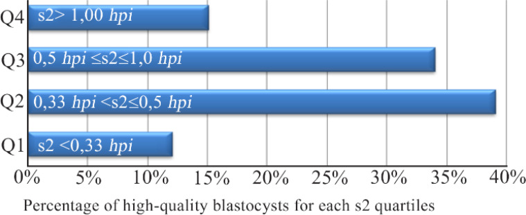
S2 median values were significantly different between high- and low-quality blastocysts (0.5 vs. 1.0 hpi, respectively; p=0.003, data not shown). The percentage of high-quality blastocysts for each s2 quartiles is displayed in figure 1. Optimal range was defined by Q2–Q3 consecutive quartiles (0.3–1.0 hr) with the highest probability of having good-quality blastocysts and significantly different values compared with other quartiles (p< 0.001).
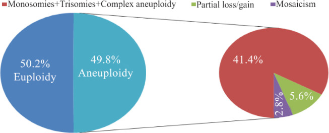
In study group B (PGT-A) (Mean age of 38.4, 95% CI of 37.5–39.3, range of 26–48), cleavage rate (No. of embryos observed on day 2/no. of normally fertilized oocytes) was 98.4% (791/804; 95% CI of 97.5–99.3). The overall blastocyst rate (No. of day 5 blastocysts/no. of normally fertilized oocytes) was 51.6% (408/791, 95% CI of 48.1–55.0) and the rate of usable blastocyst for biopsy (i.e., blastocyst with trophectoderm herniated cells) was 70.8% (289/408, 95% CI of 66.4–75.2). The overall euploid rate (No. of euploid blastocysts/no. of blastocysts ploidy results) was 50.2% (144/287; 95% CI of 44.4–56.0), while the aneuploidy rate (No. of aneuploid blastocysts/no. of blastocysts ploidy results) was 49.8% (143/287; 95% CI of 44–55.6). Among aneuploidies, 41.4% (119/287; 95% CI of 35.7–47.1) included monosomies, trisomies, and complex aneuploidies. Among these errors, a rate of 5.6% (16/287; 95% CI 2.9–8.2) was related to chromosome breakage and the effects of these alterations were partial loss or gain of chromosomal material. Finally, a small percentage (2, 8%; 95% CI of 0–4.7) was classified as mosaicism (Figure 2).
Univariate logistic regression indicated that some parameters were significantly associated with ploidy status (Table 3).
Table 3. Kinetic parameters significantly associated with development into an euploid blastocysts in a univariate logistic regression model (Group B)
| p | Odds ratio | 95.0% CI for OR | ||
|---|---|---|---|---|
|
| ||||
| Lower bond | Upper bond | |||
| tPNa | 0.010 | 1.465 | 1.291 | 1.668 |
| tPNf | 0.001 | 1.560 | 1.385 | 2.234 |
| t2 | 0.010 | 1.820 | 1.510 | 2.001 |
| t5 | 0.010 | 1.343 | 1.120 | 1.840 |
| tSB | 0.040 | 2.061 | 1.382 | 3.201 |
| s2 | 0.006 | 2.84 | 1.153 | 3.091 |
| s3 | 0.040 | 0.532 | 0.332 | 0.902 |
tPNa/tPNf: Time of pronuclei appearance/fading; t2: Time of 2-cell; t5: Time of 5-cell; tSB: Time of start of blastulation: s2: Time difference between t4 (4-cell) and t3 (3-cell); s3: Time difference between t8 (8-cell) and t5 (8-cell)
Female age was also positively associated with ploidy status (p<0.001). Using kinetic parameters as independent predictor variables of development into an euploid blastocyst and female age in a multivariate logistic regression analysis, only age (OR=1.189; 95% CI of 1.095–1.291) and timing of pronuclear appearance (tPNa) (OR=1.287; 95% CI of 1.131–1.463) predicted ploidy changes (Table 4).
Table 4. Multiple logistic regression analysis of morphokinetic parameters as predictors of development into an euploid blastocysts, adjusted for female age (Group B)
| Morphokinetic variables | p | Odds ratio | 95% CI for OR | |
|---|---|---|---|---|
|
| ||||
| Lower bound | Upper bound | |||
| Age | 0.001 | 1.189 | 1.095 | 1.291 |
| tPNa | 0.010 | 1.287 | 1.131 | 1.463 |
tPNa: Time of pronuclei appearance
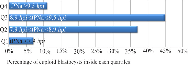
A delay in tPNa timing was observed in blastocysts that develop an aneuploidy (8.9 vs. 10.3 hpi, euploid vs. aneuploid, respectively; data not shown). Such a difference was statistically significant (p=0.03) compared with the euploid counterparts. Quartile analysis suggested that euploid rate was higher in embryos in which pronuclear appearance occurred at 7.9–9.5 hr (Q2–Q3 consecutive quartiles), as shown in figure 3. This value was statistically significant compared with the other quartiles (p<0.001).
TLM provides both undisturbed embryo culture conditions and quantitative kinetic information on development. Considering early kinetic variables, our study focused on potential implementation into clinical setting based on a comprehensive assessment of embryo viability in terms of blastocyst developmental potential and ploidy status. Present data are in line with the study hypothesis that kinetic analysis may reveal associations between cleavage patterns and embryo development to the blastocyst stage and ploidy status. Conflicting results regarding potential relationship between kinetic and ploidy status have been published (12). Establishing or even postulating a causeeffect relationship between kinetic patterns and compromised genome integrity remains elusive. Crucial nuclear events take place between appearance and fading of pronuclei, such as chromosome arrangements, histone modifications, and transcriptional activity (16). Observed differences in length of S and G2 phases may well reflect the magnitude of DNA synthesis (17). Therefore, it is possible that synthesis of altered amounts of genetic material leads to a deviation from a standard timeline of pronuclear appearance. As the factor is more strongly associated with embryo aneuploidy, advanced maternal age can play a role in the determination of kinetic patterns. Notably, maternal legacy is not restricted to the fidelity of chromosome segregation during female meiosis, but plays a role well beyond oocyte physiology, casting its influence on molecular, cellular and metabolic processes that are crucial for successful development. In our study, development to high-quality blastocyst was evaluated as one of the two endpoints. The choice to focus on development to the blastocyst stage is consistent with previous studies reporting that not only aneuploidy but also blastocyst quality is strongly associated with clinical outcome (18, 19). It was found that embryos develop more rapidly into high-quality blastocysts in complete synchrony of the second cell cycle, defined as the duration of time that embryos are at 3 cell stage, than those arrested or developed into poor quality blastocyst. Our results are in line with previous studies (20–23). As embryonic genome activation (EGA) occurs as early as the 4-cells stage, a delay or arrest in development to the blastocyst stage may reflect a defective maternal-to-zygotic transition. A recent study has shown that kinetic patterns of human fertilization can be highly predictive of embryo quality at later developmental stages (24).
The study is limited by its retrospective design and confirms that TLM cannot be adopted to predict embryo aneuploidy; however, it reveals that kinetic parameters may assist in the identification of embryos with higher ability to develop to the blastocyst stage which are less affected by chromosomal abnormalities. However, accomplishment of such a goal requires more extensive and in-depth analysis of embryo morphokinetics.
