
Perivascular Epithelioid Cell Tumors (PEC-omas) are rare mesenchymal tumors first reported in 1995. The term “PEComa” was used to describe tumors originated from perivascular epithelioid cells in 1996 (1). PEComas are a family of mesenchymal tumors including angiomyolipomas, clear cell tumors of the lung, and lymphangioleimyomas. Most of these rare tumors are benign. Besides peritoneum, the second common organ affected by the tumor is uterus (2). Up to now, about 100 patients with PEComas are reported among whom one third had uterine involvement. As affected cases of this disease were not reported, standard diagnostic and management procedure have not been described yet. In this case report, a patient with uterine PEComa is reported and described.
The patient was a 53 year old lady, who reached menopause 3 years ago, with a history of 2 vaginal deliveries. She had severe pelvic pain and abnormal uterine bleeding and underwent diagnostic curettage before visiting us. The report was normal with atrophic endometrium. Sonographic evaluation revealed a lobulated hypoechoic mass, 67×46×64 mm in size, from posterior uterine wall towards cervix. Gynaecologic evaluation revealed a mass in left side of uterus and MRI report was suggestive of a uterine sarcoma.
The patient underwent bilateral oophorectomy and hysterectomy. A 5 mm and a 12 mm trocar were used to enter abdominal cavity. A mass behind cervix and frozen pelvis were observed. An enlarged uterine with a bulging cervix, adhesion of ovaries to pelvic floor, and intestinal adhesion to cervix and left adnexa were observed. Uterine with the mass in cervix was separated intactly and removed from vagina (manipulator was not used due to probability of malignancy). The sample was sent to frozen section and in frozen section, using cryotomy and touch smear, a tumor consisting of monomorphic cells with moderate atypia and necrosis sites was observed. Mesenchymal tumor in favor of cervical sarcoma was reported. Left parametrium was thoroughly involved compressing left urethra. Right parametrium had lower involvement. Bilateral double J was inserted. The mass had sarcomatous pattern. Left parametrium was completely removed. An omental biopsy was performed. Then lymph nodes of both obturator and iliac region, left and right sides, were removed completely. An old thrombotic vein in pelvic floor and left side was observed and was not touched due to its creation of thrombosis in her last pregnancy. Samples were sent to pathology laboratory for cytological and immunohistochemistry evaluation.
In pathologic sample, a round soft mass in uterus wall, 7 cm in diameter, was observed and cervical mucosa was normal. Tissue samples were fixed by formalin 10% and colored with hematoxylineosin. A tumor consisting of monomorphic cells in regular nests and hypervascular background including delicate microvasculature separating cellular nests was observed. In larger view, multidimensional cells with clear cytoplasm and same nucleus with low to moderate atypia were observed. Cells were around vessels. Cells were colored using immunohistochemistry with HMB45, vimentin, and Melan A makers. P63, S100, cytokeratin, desmin, h-caldesmon were negative and differential diagnosis for the patient was PEC-omas (Figures 1–6).
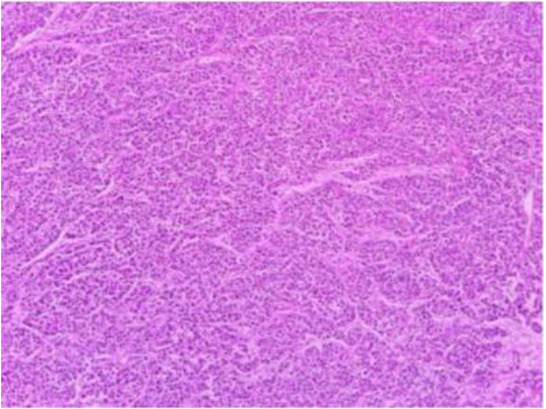
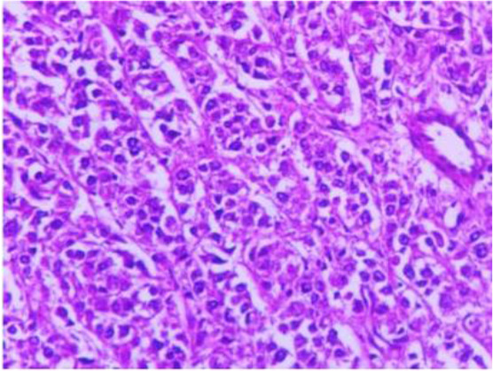

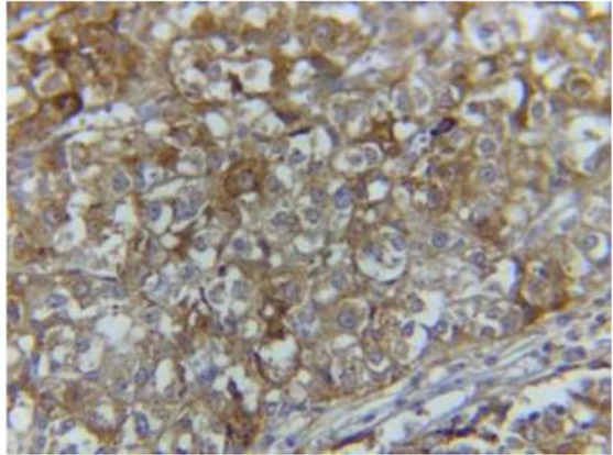
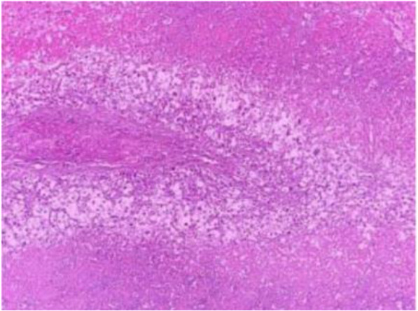
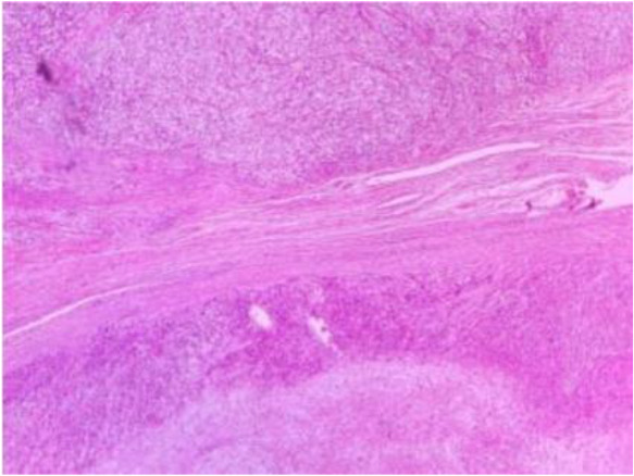
Pathology lab report revealed malignant PEC-oma without involvement of peritoneum, pelvis, parametriums, lymph nodes, and omentum. The patient was discharged after two days with a good general health and was referred to the radiotherapist. As there was no pelvic involvement, NEORAPA (antineoplastic agent) and Rapamune were prescribed for the patient. The patient is followed regularly.
Up to now, about 100 patients with PEComa of uterine are reported. Mean age of patients was about 40 years (4) though it can be observed in all age groups. Hormones may play role in its pathogenesis in women (5, 6). Malignancy potential of PEComa depends on histopharmacology of primary tumor and not its origin. PEComas are more likely to affect retroperitoneum, kindies, and female reproductive system; yet, it occurs in other body parts as well. Cervix is rarely involved (7, 8). Differential diagnosis of PEComas include all types of benign and malignant tumors. PEComas may be mistaken for carcinomas such as clear cell tumor especially if the patient has involvement of cervix or bladder (4, 9). Pathogenesis of PEComa is still unclear and it is hypothesized that they are related to complex tuberous sclerosis (9).
Clinical presentation, physical examination of pelvis, and pelvis ultrasound are not specific and can be mistaken for benign tumors or uterine fibroids or other malignancies. This non-specificity makes difficulties in diagnosis and management of PEComa and delays treatment of some patients (10–12). Furthermore, patients with malignancy have high survival rates without adjutant treatment. Chemotherapy regimen for malignant PEC-oma of reproductive system is not well defined and need more studies. Currently, the role of radiotherapy is not clear enough. Histological analysis of malignant PEComa reveals high mitotic index and multiple necrosis. Necrosis is related to tolerance threshold of chemotherapy and high angiogenesis shows high sensitivity of cells to radiation; therefore, these two statements support the role of radiotherapy for malignant PEComa (4). Systemic chemotherapy regimen without surgery is not an effective treatment. Thus, adjuvant chemotherapy has not an effective role in prognosis of patients with malignant PEComa and surgery is still the main treatment. Most malignant PEComas has good survival without recurrence or metastasis (13). Most patients with PEComa undergo bilateral oophorectomy and total hysterectomy based on their age (14). When a simple surgery may be adequate for both benign and malignant PEComa, adjuvant therapy after surgery should be provided for patients in spite of weak efficacy of radiotherapy and chemotherapy (15). Different studies have indicated increase in survival rate after surgery by using combination of chemotherapy and radiotherapy (16).
Uterine PEComa is a rare disease and the main diagnosis is made after pathologic assessment of patient’s sample following surgery and immunohistochemistry. Prognosis of the disease is depending on whether the tumor is benign or malignant. The first treatment for patients is surgery and in malignant cases, adjuvant therapy is needed for reducing the risk of recurrence and metastasis.
