
Endometriosis is a benign disease of women that causes pelvic pain, painful menstruation, and infertility (1). It is characterized by the growth of endometrial tissue outside the uterus. The ectopic cells outside the uterus act like normal endometrial tissue throughout the menstrual cycle (2). Ovaries, peritoneal cavity, and uterovesical pouch are the primary sites for the growth of ectopic endometrial tissue; however, there are rare locations, including the thorax, in which endometrial tissue grows (3). Endometriosis can affect up to 10% of women in the reproductive age, which indicates about 170 million women worldwide (4). Various methods with different sensitivities are utilized for endometriosis diagnosis, including clinical examinations, laparoscopy, imaging modalities, and laboratory tests (5). Since severe pain caused by endometriosis reduces the quality of life of patients, early diagnosis of this disease requires significant attention (6). Laparoscopy is a surgical approach for diagnosing endometriosis. Although laparoscopy is a gold standard method for endometriosis diagnosis, it is not always recommended as a first line diagnostic method (7). Medical imaging modalities, such as ultrasonography, are utilized for a less invasive assessment of endometriosis. Ultrasonographic diagnosis is often preferred over other methods due to its availability and the absence of ionizing radiation (8).
Therefore, the present study was conducted with the aim of investigating the length of the cervix and the thickness of the endometrium in women with endometriosis who were referred to the Endometriosis Clinic of Rasoul Akram Hospital in Tehran, Iran from 2018–2021.
In this case-control study, nulliparous women with and without endometriosis (diagnosed based on vaginal ultrasound) were divided into case and control groups. Before starting the study, an informed consent was obtained from the patients. The inclusion criteria consisted of healthy nulligravid Iranian women. Total number of 42 women with endometriosis were included in the case group and 42 women without endometriosis were included in the control group.
In both the case and control groups, patients underwent vaginal ultrasound, and the length and width of the cervix, the mediolateral diameter, and the thickness of the endometrium were all measured. Cervical length was meticulously evaluated using the Philips Affiniti 70 ultrasound system (software version 5.0.X; Philips, Austria) in conjunction with a RIC 5–9 MHz transvaginal probe, following bladder emptying and obtaining patient consent. Transvaginal ultrasound (TVS) was performed using a high-frequency probe of 6–7.5 MHz in the vagina (Figures 1 and 2).
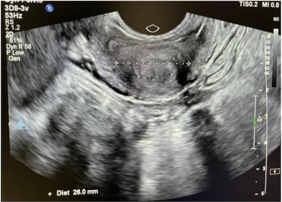
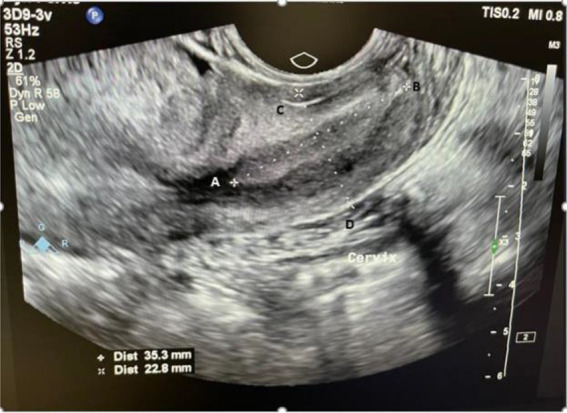
The patients of the case group underwent laparoscopy and the diagnosis was confirmed. The collected information was recorded in the registration form. Both case and control groups were matched in terms of age, weight, body mass index, age at menarche, and length of menstrual cycle. Demographic information of patients including BMI, age at menarche, height, weight, and level of education was recorded. Menstrual pain of patients was determined and recorded using visual analogue scale (VAS). VAS is a method of measuring pain using a ten-point Likert scale, ranging from 0 (no pain) to 10 (extreme pain).
The length of the cervix was measured by a radiologist using transvaginal or rectal ultrasound, from the external os to the internal os. All ultrasounds were performed on days 4 to 10 of the menstrual cycle.
Data analysis utilized descriptive statistical methods, including mean and standard deviation. The chi-square test was applied to compare the two groups when the necessary conditions were met; otherwise, Fisher’s exact test was applied.
Pearson’s correlation test was conducted to investigate the relationship between cervical length, mediolateral width, and anteroposterior length of the cervix in ultrasound imaging, along with dysmenorrhea and dyspareunia. All analyses were performed using SPSS version 22 (IBM, USA). The study was conducted in accordance with ethical guidelines, as approved by the Institutional Review Board (IR.IUMS.REC.1398.066).
A total of 84 patients were included in this study. Demographic characteristics of the patients including BMI, age, age at menarche, height, weight and level of education are shown in table 1. The average age at menarche in the control and case groups was 12.66±1.99 and 12.32±2.8 years, respectively. There was no significant difference in any of the demographic characteristics in both case and control groups (Table 1).
Table 1. Demographic characteristics of control and case groups
| Control | Case | p-value | |
|---|---|---|---|
| Age | 31.2±6.8 | 32.44±5.7 | 0.38 |
| BMI | 23.8±3.8 | 23.8±4.5 | 0.9 |
| Age at menarche | 12.66±1.99 | 12.32±2.8 | 0.5 |
| Height | 162.8±4.2 | 163.5±7.7 | 0.6 |
| Weight | 63.7±11.2 | 63.2±9 | 0.8 |
| Education level (bachelor degree and above) | 67.5% | 61.9% | 0.07 |
All patients in the case group were diagnosed with stage 3 and 4 endometrioses. In general, 23 patients in the case group (54.8%) and 27 (62.8%) patients in the control group had no dyspareunia (p=0.37). In the case group, 57% reported experiencing menstrual pain frequently or consistently, whereas 59.5% of participants in the control group indicated that they never or sometimes experienced menstrual pain. The intensity of dysmenorrhea, as measured by VAS, was 6.3±2.3 in the case group and 4.66±3.2 in the control group (p=0.01). The menstrual cycle duration was 21 to 35 days in 88.1% of the cases and 65.1% of the controls, showing significant relationship between the groups (p=0.01).
Changes of the cervical parameters in case and control groups are presented in table 2. The thickness of the endometrium in the case and control groups were 2.23±5.71 mm and 2.23±6.2 mm, respectively with no statistically significant difference (p=0.37). The anteroposterior length in the cervix was 22.54±4.36 mm in the case group and 23.33±5.1 mm in the control group (p=0.45). The average diameter of the mediolateral width (29.48±6.2 and 27.14±3.8) between the two groups was statistically significant in the endometriosis patient group and the control group, respectively (p=0.04). Cervical length was 32.39±4.26 mm in the case group and 34.19±6.5 mm in the control group, which was not statistically significant (p=0.14). The correlation between cervical length and mediolateral width measured by ultrasound was significant (r=0.4, p=0.01).
Table 2. Comparison of cervical length parameters in case and control groups
| Control | Case | p-value | |
|---|---|---|---|
| Endometrial thickness | 6.2±2.23 * | 5.71±2 | 0.37 |
| Anteroposterior length | 23.33±5.1 | 22.54±4.36 | 0.45 |
| Mediolateral width | 27.14±3.8 | 29.48±6.2 | 0.04 |
| Cervical length | 34.19±6.5 | 32.39±4.26 | 0.14 |
Millimeters
In analyzing the predictive variables related to cervical characteristics, including cervical length, mediolateral width, and posterolateral width, age and BMI of the patients were considered in predicting endometriosis. Using backward multivariate regression model that achieved an accuracy of 70%, it was found that mediolateral width had a significant directional predictive effect on endometriosis, whereas cervical length demonstrated a protective effect against the condition (Table 3).
Table 3. Variables predicting endometriosis
| ß | SE* | p-value | OR** | CI 95%*** | |
|---|---|---|---|---|---|
| Variables | |||||
| Cervical length | −0.125 | 0.058 | 0.03 | 0.88 | 0.758–0.962 |
| Mediolateral width | 0.15 | 0.061 | 0.01 | 1.17 | 1.041–1.032 |
| Constant | −0.28 | 1.68 | 0.70 |
Standard error,
Odds ratio,
Confidence interval
The purpose of this study was to investigate the length of the cervix and the thickness of the endometrium in women with endometriosis referred to the Endometriosis Clinic of Rasoul Akram Hospital in Tehran, Iran from 2018–2021. The findings of this study may enhance the diagnosis of endometriosis and contribute to a reduction in disease morbidity. It was found that demographic findings are not different in people with endometriosis and without endometriosis. The only parameter for cervical measurement obtained via transvaginal ultrasound that demonstrates a significant difference between the two groups is the mediolateral width. Also, there is a correlation between all the ultrasound indices in measuring the cervix with the indices of dysmenorrhea and dyspareunia.
Transvaginal ultrasonography is the primary method for the diagnosis of endometriosis. Ultrasonographic evaluation of cervical and endometrial indices can provide dynamic data and additional information that is not easily obtained through other imaging modalities (9).
Several studies have shown that age at menarche (10, 11), the length of menstrual cycle (12), and BMI (13) are not risk factors for endometriosis. Similarly, in the present study, there was no significant difference in age at menarche, BMI, and length of menstrual cycle in two groups, namely individuals with endometriosis and those who were healthy.
A study by Selçuk et al. (2012) demonstrated that women with endometriosis had longer menstrual cycle lengths and cervical lengths. The researchers noted that the relationship between endometriosis and demographic characteristics such as age, gravidity, parity, body mass index, menstrual cycle length, and age at menarche remains controversial. Longer cervix and thicker myometrium may be important in the etiopathology of endometriosis (14).
Research has indicated that early stages of endometriosis are often associated with junctional zone alterations in the uterus, which serve as a useful indirect marker for diagnosing the condition. According to the report of the American Society of Reproductive Medicine (ASRM), there is a strong relationship between changes in the junctional zone of the uterus and the presence of endometriosis. These findings can be obtained from ultrasonography (15, 16).
Median cervical length was greater in the control group in comparison with case group but the difference was not significant. However, in a study by Selçuk et al. (14), it was shown that the difference between the groups was significant and the endometriosis patients had longer cervical length. The difference between the groups may be attributed to the gravidity and parity of the patients in Selçuk et al.’s study, whereas our study included only nulligravid patients. The data regarding the correlation between cervical indices and endometriosis diagnosis and prognosis are quite limited. Therefore, the results of this study cannot be easily compared to the previous research.
Based on the findings of this study, mediolateral width may have a positive predictive effect on presence of endometriosis, while cervical length appears to have a protective effect against endometriosis. Further investigations with a larger sample size are required to determine the accuracy of the findings.
Our study’s findings suggest that mediolateral width in transvaginal ultrasonography can serve as a minimally invasive diagnostic tool for endometriosis. It shows a correlation with endometriosis symptoms, including dysmenorrhea and dyspareunia. Moreover, mediolateral width may have a positive predictive value for the presence of endometriosis. However, further investigations with a larger sample size are required to determine the accuracy of this study’s findings.
